Figure 2
Download original image
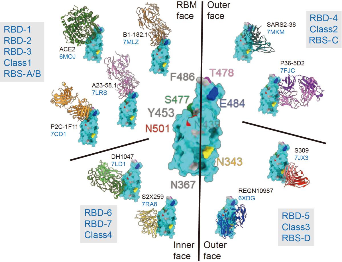
Classification of RBD-directed antibodies into seven major communities. The figure was a modification of that in Hastie et al. [52]. In the center, the entire RBD is colored in cyan together with mutations identified among the VOCs. The RRM face of RBD is depicted on the up-left corner, together with some of the representative antibodies with unknown crystal structure information. The inner and outer faces of RBD are separated by the black line in the middle. On the periphery, RBD-1 to RBD-7 communities are separated by the black lines. The relationship with previously used classification methods (Class 1–4 and RBS-A-D) are shown in grey boxes. The representative antibodies in each community are presented by their complex structures with RBD (cyan) together with their names and PDB accession numbers.
Current usage metrics show cumulative count of Article Views (full-text article views including HTML views, PDF and ePub downloads, according to the available data) and Abstracts Views on Vision4Press platform.
Data correspond to usage on the plateform after 2015. The current usage metrics is available 48-96 hours after online publication and is updated daily on week days.
Initial download of the metrics may take a while.